Home » Without Label » Back Muscles Chart : Muscles Diagrams: Diagram of muscles and anatomy charts / The teres major is a small, yet important muscle within the back.
Back Muscles Chart : Muscles Diagrams: Diagram of muscles and anatomy charts / The teres major is a small, yet important muscle within the back.
Back Muscles Chart : Muscles Diagrams: Diagram of muscles and anatomy charts / The teres major is a small, yet important muscle within the back.. Our latest youtube film is ready to run. Strain commonly occurs with incorrect lifting of heavy. Muscle spasms (contraction or stiffening of the back muscles) muscles that feel tight; The extensor muscles are attached to back of the spine and enable standing and lifting objects. 1) make midline incision along spines of vertebrae 2) extend from
It is the attachment site for the levator scapulae m. The deep back muscles, also called intrinsic or true back muscles, consist of four layers of muscles: It is an important site of muscle attachments for the intermediate layer of back muscles: We've created a free trigger point chart, which includes fybromyalgia treatment and reflexology information. Strain commonly occurs with incorrect lifting of heavy.
back anatomy chart | Workout chart, Weight training ... from i.pinimg.com Lie on your back with your knees bent and your feet flat on the floor (a). Leaning back to straight vertical and all points in between. The multifidus muscle keeps the back straight and stable. To download your free copy click the link. For more anatomy content please follow us and visit our website: The most common type of back pain is muscle pain—also called muscle strain or soft tissue strain. For images of the muscle, click on each link under location. This increases blood flow to the muscle normalizing it and bringing it back to a healthy state.
An extremely strong tendon attached to the heel.
These muscles include the large paired muscles in the lower back, called erector spinae, which help hold up the spine, and gluteal muscles. Just need a glimpse, leave your valuable advice let us know , and subscribe us! An extremely strong tendon attached to the heel. The teres major is a small, yet important muscle within the back. 1) make midline incision along spines of vertebrae 2) extend from The multifidus muscle keeps the back straight and stable. Raises and rotates arm in all directions. The trapezius is a broad, flat and triangular muscle. Three types of back muscles that help the spine function are extensors, flexors and obliques. The most common type of back pain is muscle pain—also called muscle strain or soft tissue strain. The fibres attach to the clavicle, acromion and the scapula spine. Muscle injuries of the lower back are commonly caused by an improper lift, lifting while twisting, or a sudden movement or fall, which may cause lower back pain. For images of the muscle, click on each link under location.
The trapezius and latissimus dorsi muscles connect the upper limb to the vertebral column. There are three different muscle groups found in the back: Our latest youtube film is ready to run. 1) make midline incision along spines of vertebrae 2) extend from These structures work together to support the body, enable a range of movements, and send messages from the.
Pin em Muscle from i.pinimg.com Superficial, intermediate, deep and deepest layers.these muscles lie on each side of the vertebral column, deep to the thoracolumbar fascia they span the entire length of the vertebral column, extending from the cranium to the pelvis The most common causes of lower back pain are strain and problems with back structures. To download your free copy click the link. The back's muscles start at the top of the back (named the cervical vertebrae) and go to the tailbone (also named the coccyx). Most of the time, back muscle pain is diagnosed then treated with little more than a prescription of rest, painkillers and muscle relaxants. We are pleased to provide you with the picture named anatomy of back muscles diagram.we hope this picture anatomy of back muscles diagram can help you study and research. An extremely strong tendon attached to the heel. This procedure is one of the most powerful yet simple ways to treat muscle pain and discomfort.
Lie on your back with your knees bent and your feet flat on the floor (a).
For images of the muscle, click on each link under location. 1) make midline incision along spines of vertebrae 2) extend from The intermediate layer contains the erector spinae muscles, whose many functions include the extension and lateral flexion of the spine, head and neck. Claim your free copy of the client back care guide today. If you experience any of these symptoms, seek medical attention immediately. It is the attachment site for the levator scapulae m. The muscles of the lower back help stabilize, rotate, flex, and extend the spinal column, which is a bony tower of 24 vertebrae that gives the body structure and houses the spinal cord.the spinal. The muscles on each side form a trapezoid shape. Strained muscles often cause back pain. Other muscles are small and cover much less space. The back isn't only one of the body's biggest and strongest body parts, it's also the most complicated in terms of being a series of interconnected muscle groups. Anatomy chart courtesy of fcit the latissimus dorsi muscles (also known as the lats) are the largest muscles of the back. Strain commonly occurs with incorrect lifting of heavy.
The achilles tendon in the strongest in the body. Anatomy chart courtesy of fcit the latissimus dorsi muscles (also known as the lats) are the largest muscles of the back. Our latest youtube film is ready to run. Multifidus issues usually lead to other problems due to improper recruitment of other muscles to avoid pain. The extensor muscles are attached to back of the spine and enable standing and lifting objects.
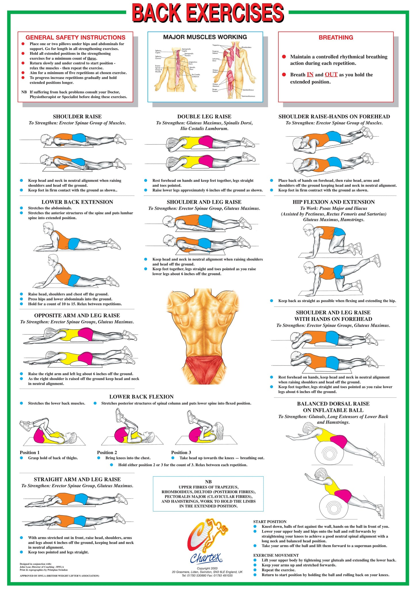
Back Muscles Floor Exercise Chart - Chartex Ltd from cdn.shopify.com The superficial group, the deep group, and the intermediate group. These structures work together to support the body, enable a range of movements, and send messages from the. Three types of back muscles that help the spine function are extensors, flexors and obliques. Our latest youtube film is ready to run. This increases blood flow to the muscle normalizing it and bringing it back to a healthy state. Extends spine and trunk back. Raises and rotates arm in all directions. Artery) p.134 accessory nerve p.
This procedure is one of the most powerful yet simple ways to treat muscle pain and discomfort.
Muscle charts of the human body for your reference value these charts show the major superficial and deep muscles of the human body. Three types of back muscles that help the spine function are extensors, flexors and obliques. Certain back muscles extend to other areas, like the shoulders, upper arms, and thighs. The back isn't only one of the body's biggest and strongest body parts, it's also the most complicated in terms of being a series of interconnected muscle groups. We are pleased to provide you with the picture named anatomy of back muscles diagram.we hope this picture anatomy of back muscles diagram can help you study and research. An extremely strong tendon attached to the heel. A strain can be an injury to a tendon attachment from muscle to bone. The achilles tendon in the strongest in the body. Try these exercises to stretch and strengthen your back and supporting muscles. These muscles include the large paired muscles in the lower back, called erector spinae, which help hold up the spine, and gluteal muscles. The most common type of back pain is muscle pain—also called muscle strain or soft tissue strain. For images of the muscle, click on each link under location. It is the most superficial of all the back muscles.